Keratosis Is a Thickened Area of the Epidermis
A macule is a discoloration of the skin that is flat and level with the skin. The area beneath the free edge of the nail furthest from the cuticle is called the hyponychium.
Skin And Lacrimal Drainage System Ento Key
A tender thickened ulcerated or enlarging actinic keratosis is suspicious of evolution to SCC.

. The keratinocyte is the most common type of skin cell in the epidermis the. Being able to recognize the early signs of actinic keratosis is important because these precancers can be treated and removed before they turn into cancer. The layer of the skin above the dermis is the.
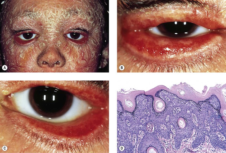
Cutaneous horn may arise from an underlying actinic keratosis or SCC. 5 Lesions present in a patient in the sixth to eighth decade of life as a pigmented and thickened area of pigment on the lower eyelid with irregular borders. Actinic Keratosis also known as solar keratosis is a scaly or crusty lesion on the skin that develops slowly and indicates the presence of sun damage.
Discoid lupus erythematosus is the most common type of chronic cutaneous lupus CCLE an autoimmune skin condition on the lupus erythematosus spectrum of illnesses. 1018 In any patient with a pigmented lesion biopsy should be considered. The color of actinic keratosis will depend on your skin tone and may look pink red dark tan white or the color of your skin.
Actinic keratosis improves just two days after a. This is the outermost layer of the skin visible to the eye. Because they are sun damaged people with actinic keratoses are also at risk of developing actinic cheilitis basal cell carcinoma BCC which is more common than SCC melanoma and rare forms of skin cancer.
10818Additional findings include focal scale andor crusts a thin rolled border and variable amounts of. It consists of a thickened layer of stratum corneum. Crusty pre-cancerous growths caused by.
Epidermis may be hyperplastic and papillomatous and may have keratotic cysts and pigment in basal epidermal keratinocytes Loose fibrocollagenous stroma abundant vessels Usually no adnexa Variable adipose tissue in larger ones - lipofibroma Traumatic changes. Lichen simplex chronicus epidermal necrosis ulceration pagetoid dyskeratosis lichen sclerosus-like. The most common form of skin cancer that strikes in the top layer of the epidermis moles.
Basal cell carcinoma. The two main types of human skin are glabrous skin the nonhairy skin on the palms and soles also referred to as the palmoplantar surfaces and hair-bearing skin. The term keratosis refers to a knobby overgrowth of keratinocytes.
Actinic keratosis usually appears as a patch of dry scaly skin. Keratosis is an area of the skin that is overgrown or thickened. Melanoma of the eyelid is a relatively rare tumor making up less than 1 percent of eyelid cancers.
Within the latter type hairs in structures called. The epidermis is composed of multiple layers of. It is most commonly found on parts of the body frequently exposed to the sun including the bald scalp face ears lips backs of the hands or forearms neck and shoulders.
Melano pertains to. The skin is composed of three layers. The nail is an accessory structure of the integumentary system.
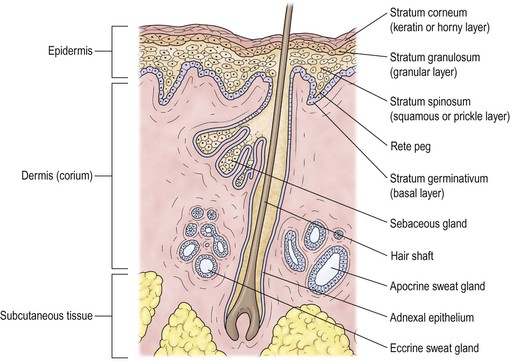
Dark growths on the skin actinic keratosis. It presents with red painful inflamed and coin-shaped patches of skin with a scaly and crusty appearance most often on the scalp cheeks and ears. The epidermis the dermis composed of the superficial papillary and deeper reticular dermis and the hypodermis.
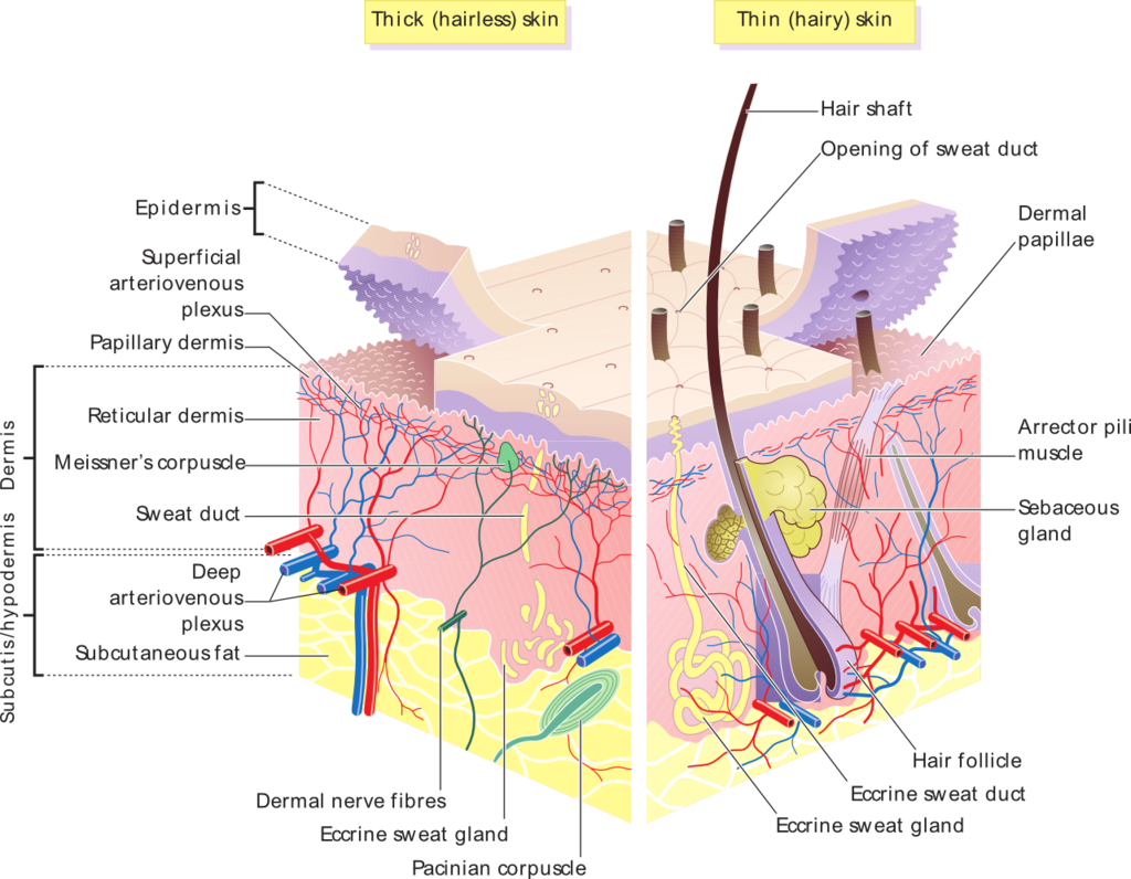
The skin has structural differences among the different areas of the body in terms of epidermal and dermal thickness distribution of appendages and pigmentation. Squamous cell carcinoma SCC also known as squamous cell cancer is the second most common type of skin cancer following basal cell carcinomaAbout 1 million cases are diagnosed each year in the United States. 615 The usual actinic keratosis is characterized by focal parakeratosis with loss of the underlying granular layer and a slightly thickened epidermis with some irregular downward buds Fig.
Superficial BCCs typically present as a well-circumscribed erythematous maculepatch or thin papuleplaque with the diameter varying from a few millimeters to several centimeters Fig. Bolognia MD in Dermatology 2018 Superficial basal cell carcinoma. Diagnostic biopsy is undertaken in only a small percentage of actinic keratoses diagnosed clinically.
A seborrheic keratosis is a type of skin growth. The skin weighs an average of 4 kg 88 lb covers an area of 2 m 2 22 sq ft and is made of three distinct layers. It begins in the squamous skin cells located in the top layer of skin called the epidermisThe DNA in squamous cells can become damaged from.
545 The clinical accuracy in the recognition of actinic keratoses varies from 74 to 94. Actinic keratosis is thickening of the outer layer of the skin caused by prolonged exposure to sunlight. It contains specialized cells responsible for pigmentation of the skin melanocytes protecting the skin Langerhans cells and allowing the skin to feel pressure Merkel cells.
It contains a network of tough but elastic collagen fibers that make the skin. The epidermis dermis and subcutaneous tissue. This is the middle layer of the skin.
Keratosis Actinic Actinic means ray or radiation.

Layers Of The Skin Anatomy And Physiology I

An Acanthotic Type Of Seborrheic Keratosis Taken From The Left Download Scientific Diagram

Chapter 16 Flashcards Quizlet

Stratum Corneum Top Layer Of Skin Anatomy And Function
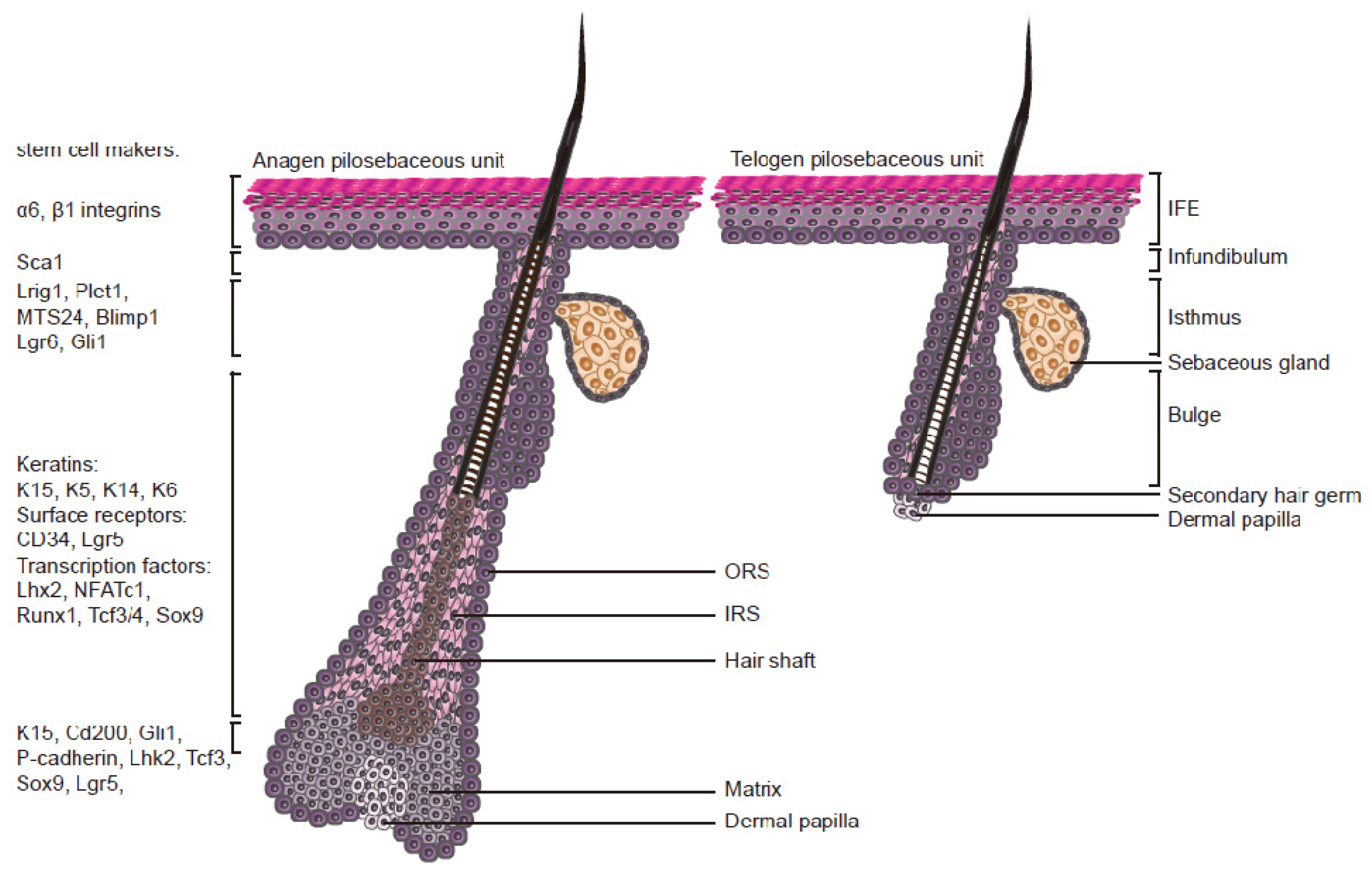
Ijms Free Full Text Egfr Ras Raf Signaling In Epidermal Stem Cells Roles In Hair Follicle Development Regeneration Tissue Remodeling And Epidermal Cancers Html

A Skin Was Thickened Black Arrows Showing Two Small Elevated Masses Download Scientific Diagram
![]()
Skin Cells Layers And Histological Features Kenhub

F Skin The Integument Diagram Quizlet
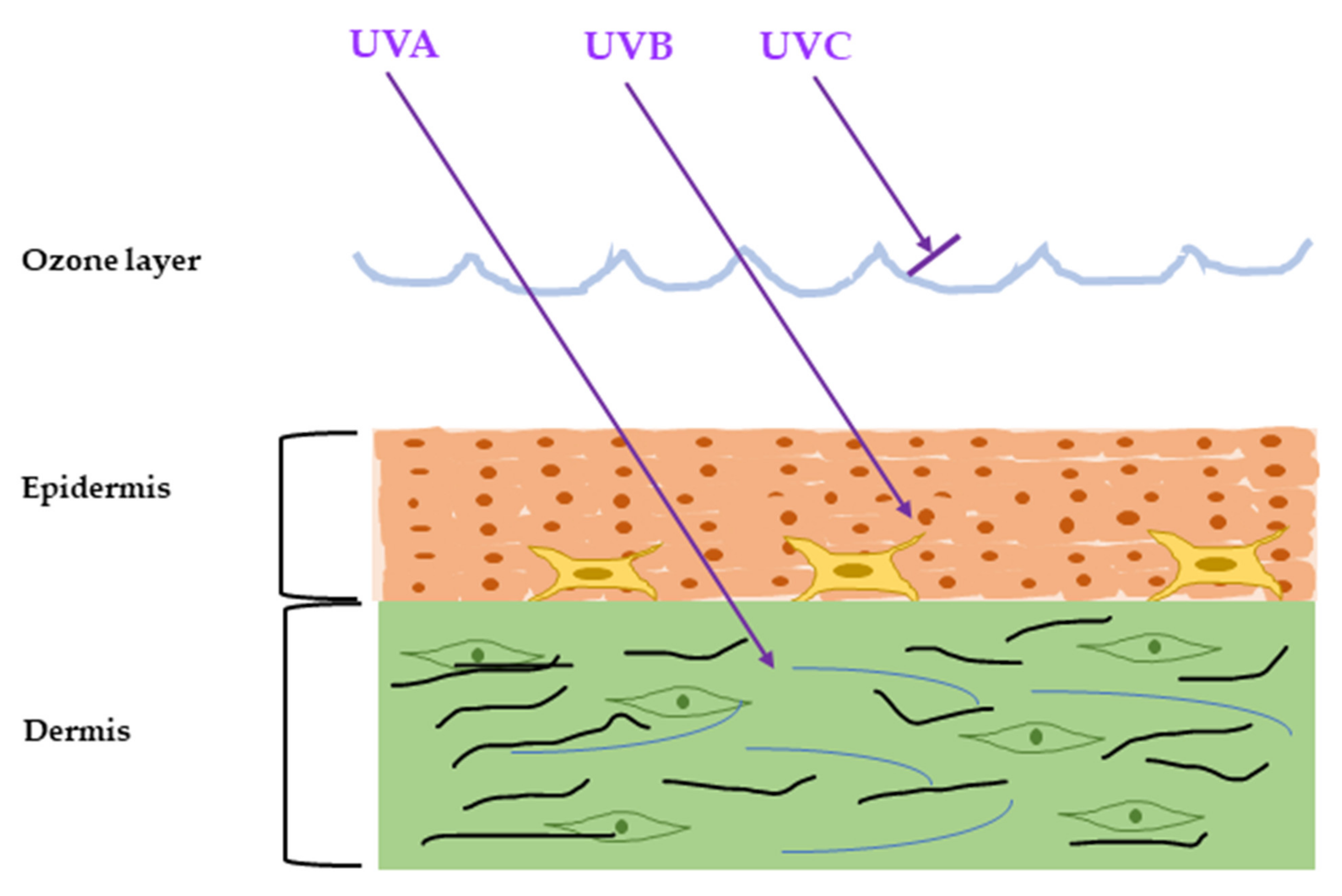
Ijms Free Full Text Inflammatory Molecules Associated With Ultraviolet Radiation Mediated Skin Aging Html

Layers Of The Skin Anatomy And Physiology I

Usmle Step 1 Qbank Skin Pathology Dermatology Nurse Pharmacology Nursing Pathophysiology Nursing

Pin On Skinpedia Jo

Histology Displays An Area Of Hypokeratosis Demarcated By A Sharp And Download Scientific Diagram
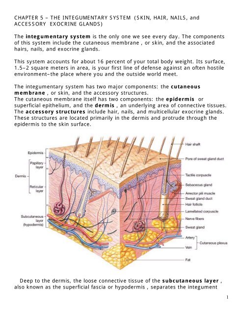
Chapter 5 The Integumentary System Skin Hair
The Skin Boundless Anatomy And Physiology

The Pathobiology Of Skin Aging The American Journal Of Pathology
Skin And Lacrimal Drainage System Ento Key

Ch 14 Skin Flashcards Quizlet
Skin And Lacrimal Drainage System Ento Key


Comments
Post a Comment